
Sinboda opens a new window into single-molecule conformational analysis.
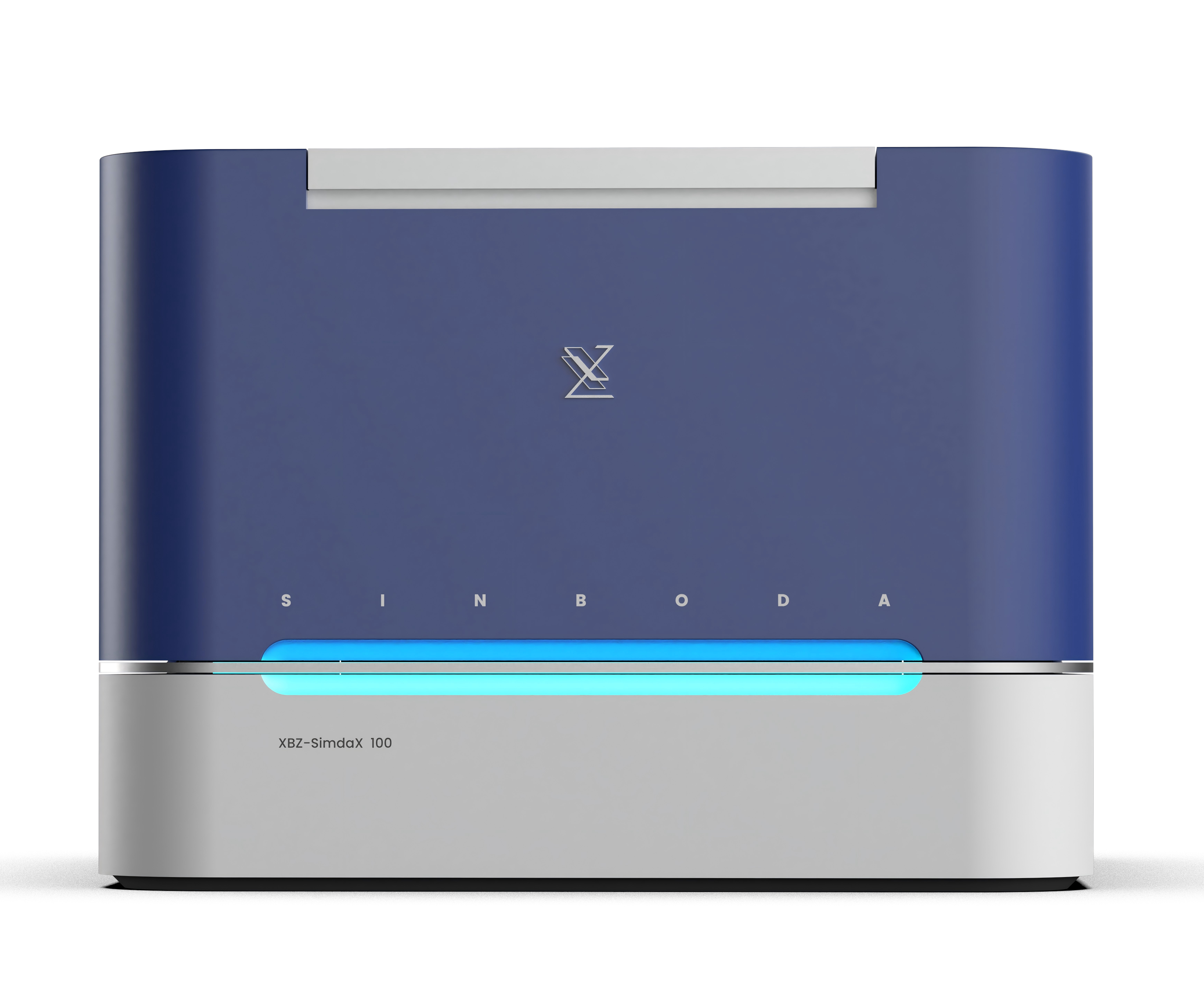
XBZ-SimdaX 100 by Sinboda is the first ultra-sensitive, visualized protein detection platform based on single-molecule imaging. It enables simultaneous quantitative and morphological characterization of specific protein molecules in samples such as plasma, serum, and cerebrospinal fluid, providing solutions for early disease screening, diagnostic assistance, companion diagnostics, therapeutic evaluation, drug development, and novel biomarker validation to meet diverse scientific and clinical needs.

Achieves single-molecule level capture and visualization

Detection limit as low as fg/mL

Differentiating monomers and aggregates—the degree of aggregation serves as an important indicator of disease progression;
Through specialized experimental design, conformational changes in proteins can be detected.

Measures aggregation degree, distribution, abundance, and total concentration of target proteins in blood samples, achieving integrated quantitative and morphological characterization and providing multidimensional analytical validation for research and clinical applications.
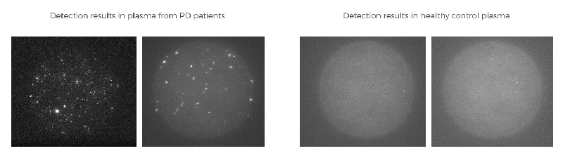
Proteins such as Aβ, Tau, and α-synuclein are prone to self-aggregation due to their intrinsic β-sheet structures. The morphological differences between monomers and aggregates (size, shape, brightness) are distinct and measurable. At the same concentration, the degree of aggregation varies significantly. These morphological differences are crucial indicators of neurodegenerative disease progression. Using highly specific antibodies for molecular capture and labeling, high-resolution single-molecule imaging enables combined quantitative and morphological characterization.
High-resolution single-molecule imaging enables signal identification at the level of individual fluorescent molecules, allowing more precise quantification. Differences in the number of fluorescent molecules associated with monomers and aggregates lead to variations in brightness and size. AI algorithms detect spots of varying intensity and size, identifying proteins at different aggregation states. This allows multidimensional characterization of aggregation degree, distribution, abundance, and total concentration, enabling more accurate early screening and auxiliary diagnosis of neurodegenerative diseases.